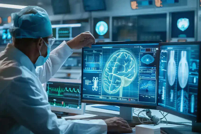
In the rapidly evolving world of healthcare, medical imaging technology stands as a cornerstone of modern diagnostics and treatment. Over the years, these technologies have advanced by leaps and bounds, transforming how we understand, diagnose, and treat various medical conditions. From the early days of X-rays to the current innovations in AI-powered imaging, the journey of medical imaging is both fascinating and crucial for the future of medicine.
Evolution of Medical Imaging Technology
Early Stages of Medical Imaging
The journey of medical imaging technology began with the landmark discovery of X-rays by Wilhelm Conrad Roentgen in 1895. This breakthrough allowed doctors to look inside the human body without making any incisions, a revolutionary development in medical science at the time. X-rays quickly became a fundamental diagnostic tool, offering invaluable insights into a variety of conditions, particularly those involving bone fractures, dental issues, and lung problems. The ability to non-invasively visualize internal structures changed the way physicians approached diagnosis and treatment, leading to more accurate and timely interventions. The widespread adoption of X-rays marked the beginning of a new era in medicine, where visualization of the body’s internal workings became a key component of medical care.
The Rise of Advanced Imaging Techniques
The evolution of medical imaging took another significant leap forward with the introduction of Computed Tomography (CT) scans in the 1970s. CT scans combine the principles of X-rays with computer processing to create detailed cross-sectional images of the body. This technology allows for a comprehensive examination of internal organs, blood vessels, and bones, providing more detailed information than a standard X-ray. CT scans revolutionized the field by enabling earlier and more accurate diagnoses of conditions such as cancer, cardiovascular diseases, and traumatic injuries. The ability to visualize the body’s interior in such detail has made CT scans an indispensable tool in emergency medicine, oncology, and surgery, among other fields.
Magnetic Resonance Imaging (MRI) further transformed medical imaging by offering an entirely new way to visualize soft tissues within the body. Introduced in the late 20th century, MRI uses strong magnetic fields and radio waves to produce detailed images, without the use of ionizing radiation. This technology quickly became the gold standard for diagnosing a wide range of conditions related to the brain, spinal cord, and musculoskeletal system. MRI’s ability to provide high-contrast images of soft tissues made it particularly useful in detecting tumors, brain injuries, and other complex conditions that were difficult to assess with previous imaging methods. The precision and clarity of MRI have made it a cornerstone of modern diagnostic medicine, enabling more effective treatment planning and patient care.
Breakthroughs in Medical Imaging
PET and SPECT Scans
Positron Emission Tomography (PET) and Single Photon Emission Computed Tomography (SPECT) have marked significant advancements in the realm of nuclear medicine. Unlike traditional imaging methods that primarily capture the structural aspects of the body, PET and SPECT scans delve deeper into the metabolic processes occurring within the body’s tissues. This capability is essential for diagnosing and monitoring a range of complex conditions, including:
- Cancer: PET scans are particularly effective in detecting areas of high metabolic activity, which often correlate with cancerous cells. This makes PET scans a vital tool in oncology for both initial diagnosis and the assessment of treatment effectiveness.
- Heart Disease: SPECT scans are frequently used to evaluate blood flow and heart function, helping in the diagnosis of coronary artery disease and other cardiovascular conditions.
- Neurological Disorders: Both PET and SPECT scans provide crucial insights into brain activity, aiding in the diagnosis of conditions such as Alzheimer’s disease, epilepsy, and Parkinson’s disease by mapping functional changes in the brain.
These imaging techniques provide a functional map of the body, offering insights that go beyond what structural imaging can provide. This is particularly valuable in complex cases where understanding the underlying metabolic or functional abnormalities is key to developing effective treatment plans.
3D and 4D Imaging
The introduction of 3D imaging has revolutionized the way medical professionals visualize the human body, offering a more comprehensive view of its complex structures. This technology is especially beneficial in areas that require detailed anatomical analysis, such as:
- Orthopedics: 3D imaging allows for precise visualization of bones and joints, aiding in the diagnosis and treatment planning for fractures, deformities, and other musculoskeletal conditions.
- Surgical Planning: Surgeons use 3D models to navigate intricate anatomical structures with greater accuracy, which improves surgical outcomes and reduces the likelihood of complications.
Building upon the capabilities of 3D imaging, 4D imaging introduces the element of time, allowing for the real-time visualization of moving structures within the body. This advancement is particularly useful in:
- Cardiovascular Diagnostics: 4D imaging captures the dynamic movements of the heart and blood flow, providing crucial information for diagnosing heart conditions and planning interventions.
- Obstetrics: In prenatal care, 4D imaging enables real-time monitoring of fetal movements and development, offering a more detailed assessment of the pregnancy.
These breakthroughs in imaging technology not only enhance diagnostic accuracy but also provide valuable tools for treatment planning and monitoring, leading to better patient outcomes across various medical fields.
The Role of AI and Machine Learning in Medical Imaging
AI-Powered Image Analysis
Artificial Intelligence (AI) and Machine Learning (ML) are revolutionizing the field of medical imaging by introducing unprecedented levels of speed and accuracy in image analysis. AI-powered tools are capable of processing vast amounts of imaging data in a fraction of the time it would take a human radiologist. These systems are designed to identify patterns and anomalies that may be too subtle or complex for the human eye to detect, thereby enhancing the diagnostic accuracy significantly. This ability is particularly beneficial in detecting early-stage diseases, where minor changes in imaging data can indicate the onset of serious conditions such as cancer. As a result, AI-driven image analysis is becoming an indispensable tool in the early detection and diagnosis of various diseases.
Beyond enhancing accuracy, AI also plays a critical role in reducing human error in medical imaging. The automation of the image analysis process through AI systems provides an additional layer of scrutiny, helping to catch potential mistakes that might be overlooked by human reviewers. This dual approach ensures that patients receive the most accurate diagnosis possible, which is crucial for effective treatment planning. Furthermore, the use of AI in medical imaging significantly reduces the time required to interpret imaging results, enabling faster decision-making in clinical settings, which can be particularly important in emergency situations.
| Aspect | Traditional Imaging | AI-Powered Imaging | Benefits of AI |
| Speed of Analysis | Time-consuming | Extremely fast | Reduces diagnostic time significantly |
| Accuracy | Prone to human error | High precision | Improves diagnostic accuracy |
| Error Detection | Manual review | Automated review | Minimizes human error, provides second review |
| Early Disease Detection | Dependent on expertise | Pattern recognition | Detects diseases at earlier stages |
Predictive Analytics and Imaging
The predictive capabilities of AI are transforming the landscape of disease detection and prevention. By leveraging large datasets of medical images, AI algorithms can identify early signs of diseases such as cancer, often before symptoms manifest. This early detection is crucial for improving patient outcomes, as it allows for timely interventions that can significantly increase survival rates. In addition to early detection, AI also enables continuous monitoring of patients, providing real-time analysis that can alert healthcare providers to changes in a patient’s condition that may warrant immediate attention.
AI’s impact extends beyond detection into the realm of personalized medicine. By integrating imaging data with other patient information, such as genetic profiles and medical history, AI can help develop highly personalized treatment plans. These tailored strategies are designed to maximize the effectiveness of treatment while minimizing potential side effects, offering a more precise approach to healthcare. For example, in oncology, AI can analyze imaging data alongside genetic information to suggest the most effective chemotherapy regimen for a particular patient, thereby enhancing treatment outcomes.
| Aspect | Traditional Approach | AI-Enhanced Approach | Benefits of AI |
| Early Disease Detection | Reactive, symptom-based | Proactive, data-driven | Enables earlier intervention, improves outcomes |
| Personalized Treatment Plans | Generalized approach | Tailored to individual | Increases treatment effectiveness, reduces side effects |
Integration of Medical Imaging with Other Technologies
Fusion Imaging
Fusion imaging is an advanced technique that integrates different imaging modalities, such as Positron Emission Tomography (PET) and Computed Tomography (CT), to provide a more comprehensive view of the body’s internal structures. By combining these modalities, fusion imaging allows healthcare providers to correlate anatomical details with functional information, leading to more accurate and informed diagnoses. This approach is particularly beneficial in complex cases where a single imaging modality may not provide enough information. For instance, while a CT scan might reveal the precise location and size of a tumor, a PET scan can show the metabolic activity within that tumor, providing a fuller picture of its behavior and potential aggressiveness.
In oncology, fusion imaging has become a critical tool in assessing the extent of cancer and planning treatment strategies. The ability to merge detailed anatomical images from a CT scan with the metabolic data from a PET scan allows doctors to better understand the tumor’s behavior, such as its growth rate and response to therapy. This comprehensive view is essential for developing effective treatment plans, which may include surgery, radiation, or chemotherapy. By providing a more complete picture of the disease, fusion imaging enables more precise targeting of cancerous tissues while sparing healthy surrounding tissues, ultimately improving patient outcomes.
| Imaging Modality | Primary Use | Advantages in Fusion Imaging | Applications |
| CT Scan | Anatomical detail | Provides detailed structural images | Tumor localization, surgical planning |
| PET Scan | Metabolic activity | Reveals functional information of tissues | Cancer detection, treatment response |
| Fusion Imaging | Combined analysis | Correlates structure and function | Comprehensive cancer assessment, complex cases |
Wearable Imaging Devices
Wearable imaging devices represent a significant innovation in medical technology, offering continuous and real-time monitoring of patients outside traditional healthcare settings. These devices, which can be worn like a wristwatch or patch, continuously collect imaging data that can be used to monitor the progression of diseases or the effects of treatment. This real-time data collection is particularly valuable for managing chronic conditions, where constant monitoring can help detect early signs of deterioration or complications, allowing for timely interventions. For instance, in cardiology, wearable devices that monitor heart activity can alert doctors to arrhythmias or other cardiac events before they become life-threatening.
The portability of wearable imaging devices also makes them ideal for use in remote or underserved areas, where access to traditional imaging facilities may be limited. These devices offer a promising solution for bringing advanced medical imaging to populations that might otherwise be underserved, improving healthcare access and equity. Additionally, the ability to monitor patients continuously without the need for hospital visits can reduce healthcare costs and increase patient comfort, making wearable imaging devices a valuable tool in modern healthcare.

